Shining a Light on Dental X-Rays
Dental x-rays are a valuable part of your dental treatment. They allow your dentist the ability to more effectively evaluate your oral health and detect damage or other conditions that may not be visible during an oral examination. For example, x-rays can show the condition of your teeth and roots. They can show the presence of cavities, abscesses, impacted teeth, or teeth that have not fully developed as well as abnormal growths such as cysts and tumors.
The most common type of x-ray is the bitewing x-ray. This requires the patient to hold, or bite down on a piece of plastic with x-ray film in the center.
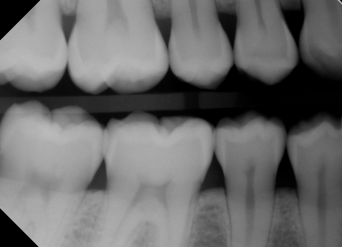
These x-rays are used to check for decay between the teeth, one of the most common areas for bacteria to reside. They show how well the upper and lower teeth line up, indicate bone loss and help determine if an infection is present. Typically these x-rays are taken once a year, or as needed.
Another type of x-ray is the panoramic x-ray.
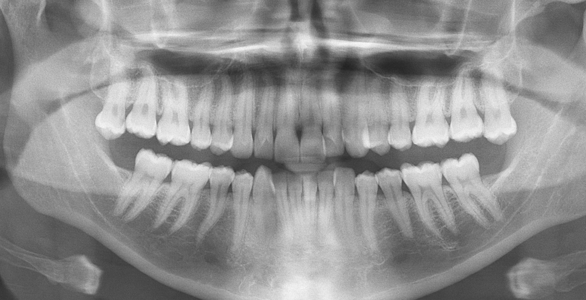
Just like a photographer can capture a wide angle view with a panoramic photograph, the panoramic x-ray allow dentists to see the entire structure of your mouth in one image. It gives a full view of the oral cavity including the jaw and joints, sinuses and nasal area. A common use of a panoramic x-ray is to assess teeth development in a child or teenager, especially wisdom teeth. Often, the wisdom teeth don’t erupt until the teenage years or later, possibly causing them to become crowded or impacted (when they do not have enough room to grow). The panoramic x-ray can also evaluate the progression of TMJ, expose cysts and abnormalities and help treatment plan for dentures, braces and implants. Panoramic x-rays are simple to perform and unlike bitewings, the x-ray film is inside a mechanism that rotates around your head. Typically these x-rays are done every 5 years or as needed.
With the use of this technology, it does raise the concern about safety and exposure to radiation. Recently a study published in Radiation Protection Dosimetry concluded that the amount of radiation a patient is exposed to during a dental x-ray is a fraction of the natural daily background exposure and that hypothetical cancer risks are speculative and should be discouraged.
For more information, see the full study, “Optimizing Radiographic Bitewing Examination to Adult and Juvenile Patients Through the use of Anthropomorphic Phantoms, published in Radiation Protection Dosimetry online first Aug. 4, 2013.
Leave a reply →